User login
From Pharma’s Factories Direct to You
Pharmaceutical giant Eli Lilly recently announced that its newly approved weight loss medication Zepbound — a glucagon-like peptide 1 receptor agonist (GLP-1 RA) akin to Mounjaro, Ozempic, and Wegovy — will be prescribed by independent telehealth providers on a platform managed by the company itself. The drug can be subsequently shipped direct to consumer (DTC), allowing delivery straight to patients’ homes.
This arrangement raises serious concerns about an inherent conflict of interest, as we previously discussed. What happens when a pharmaceutical company influences access to remote providers who prescribe the very medications it manufactures?
Without new guardrails, the potential for misleading messaging to result in dangerous prescribing patterns looms large. The United States is one of only two countries to allow DTC advertising of prescription drugs, and the explosion in demand for GLP-1 RAs is partly attributable to this model (Oh, oh, Ozempic, anyone?). Americans spent over $78 billion on weight loss goods and services in 2019; time-intensive approaches such as diet and exercise are understandably difficult, and the public has always looked for a magic cure. Although GLP-1 RAs are promising, they may present a path to disaster without proper supervision.
LillyDirect, which in addition to Zepbound offers migraine medications and other products in the company’s catalogue, primarily aims to increase access to medication and reduce costs of the drugs for consumers. The stated mission is noble: By cutting out the middlemen of traditional pharmacies and benefit managers, administrative costs drop. LillyDirect goes a step further by reducing the need for patients to visit their regular family doctor to receive these medications.
On the surface, this design appears promising. Wait times for doctor’s appointments will fall. Patients can order drugs from the comfort of their home. Everyone benefits. Or do they?
Although easier access and reduced cost may be an apparent win for patients, DTC arrangements complicate the ethics of prescriptions and patient follow-up. This model reminds us of the roots of the opioid crisis, where powerful advertising and relationships between prescribers and drugmakers led to great harm. Providers often faced a conflict of interest when prescribing dangerous drugs to patients who requested them. We must learn from these mistakes to ensure there is critical oversight into the independence of prescribers used by LillyDirect and other DTC platforms.
Adding to these parallels, once a patient begins a GLP-1 medication such as Zepbound, stopping treatment will probably lead to regaining lost weight, serving as negative reinforcement. Hence, patients may decide never to discontinue these medications.
Obtaining what amounts to a lifelong prescription from a telehealth provider who may never follow a patient sets a dangerous precedent that will be difficult to unravel once begun. Recent challenges in access to medications such as Zepbound have been complicated by supply chain and manufacturing issues, leading to potential interruptions in patient access, ultimately affecting compliance. The rapid increase in online providers indicates competition for distribution channels has sharply increased and poses a threat to Lilly’s DTC site.
Furthermore, the lack of a regular physician to monitor patients introduces uncertainty in safety and continuity of care. These are important tenets in protecting patients, especially patients who are not diabetic and desire a quick fix. We have already seen a huge, arguably unrestrained, rise in prescriptions of GLP-1 RAs for weight loss — up to a 352% increase in 2023.
These drugs have shown great promise and are generally safe when used in the right patient, but important contraindications exist — namely, serious gastrointestinal side effects and low blood glucose in nondiabetic persons — that an astute physician must consider. Patients desiring these medications often must undergo comprehensive laboratory testing and cardiac evaluation, both before initiation and during regular follow-up, to check for comorbidities.
The American College of Physicians cautioned against such prescribing practices in a recent position statement, emphasizing that the lack of an established care provider could adversely affect patients. We note that the potential harms of DTC sales would concentrate in economically and racially underserved communities, where obesity, lack of insurance, and low health literacy are more common.
But the DTC genie is out of the pill bottle, and as such platforms become more common, patients will inherently take more ownership over their medical care. Remote providers will of course not be following these patients and evaluating for side effects. As a result, we in medical practice must be abreast of new downsides of these medications if and when they arise.
Every clinician must be aware of the medications a patient is taking, even those that they did not prescribe. They should educate their patients about drug-drug interactions and side effects and order lab tests to monitor for side effects.
Independent physicians abide by an underlying oath: First, do no harm. They serve as a trusted check on industry and a valuable long-term partner for patients. Where are the guardrails to protect patients and ensure that pharmaceutical companies are not essentially pushing prescriptions for their own products? Will traditional healthcare providers be effectively relegated to a bystander role in Lilly’s transactional approach to medication distribution? Unlike other commercial goods, pharmacologics have great nuance; not every approved medication is meant for every patient.
A version of this article appeared on Medscape.com.
Knee Osteoarthritis Trials Show Promising Results for Several Novel Injectables
VIENNA — Encouraging primary or secondary analyses of trial data for the use of several novel injectables and gene therapy for knee osteoarthritis (OA) were reported at the OARSI 2024 World Congress.
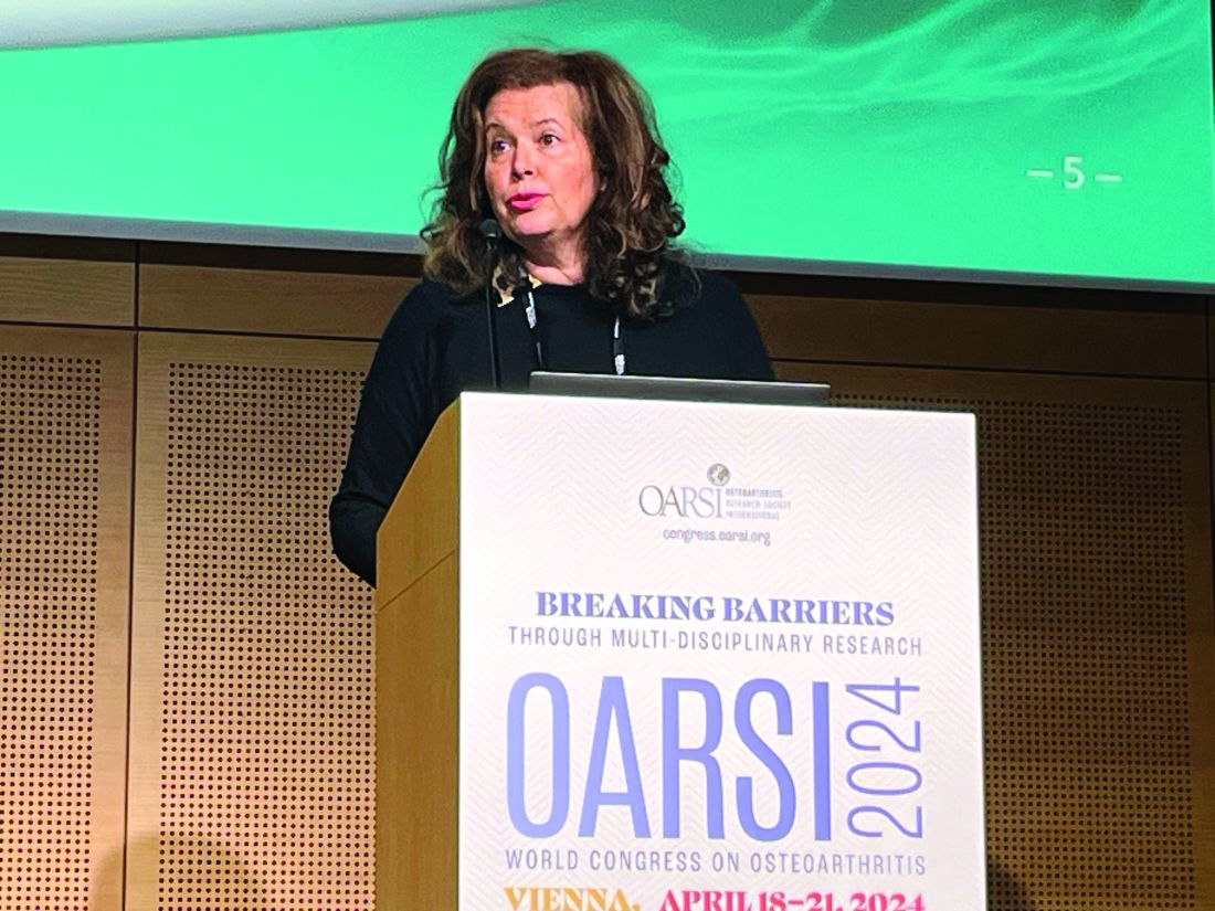
Of all the approaches discussed during the News in Therapies session at OARSI 2024, the most intriguing was the use of the placental extract PTP-001 (MOTYS, Bioventus), session chair Nancy E. Lane, MD, of the University of California Davis School of Medicine, Sacramento, California, told this news organization.
Other notable presentations of data from trials of investigational agents for knee OA included an update from the SPRINGBOARD phase 2B trial of EP-104IAR, a novel long-acting formulation of the corticosteroid fluticasone propionate; a phase 2 trial of pentosan polysulfate sodium (PPS), a non-opioid, semi-synthetic xylose-based polysaccharide; and an update on phase 2 study results for XT-150, a non-viral, plasmid-based gene therapy designed to express a proprietary variant of interleukin 10 (IL-10).
PTP-001 (MOTYS)
Indeed, promising results were seen in a phase 2 trial testing a single intra-articular (IA) injection of PTP-001 vs an IA saline placebo in just over 200 individuals with symptomatic knee OA. Results of this dose-finding study were presented by Alessandra Pavesio, senior vice president and the chief science officer of Bioventus/Doron Therapeutics, Durham, North Carolina.
Ms. Pavesio reported there were decreases in knee pain and improvements in knee function, as measured using the Western Ontario and McMaster Universities Arthritis Index (WOMAC). These changes were seen after 26 weeks of treatment with PTP-001 given at either a low (100 mg, n = 74) or high (200 mg, n = 40) dose.
Although the changes were only numerically and not statistically different from placebo (n = 71) when looking at the total study population, Ms. Pavesio noted that a key objective of the trial had been to identify populations of patients that may benefit.
When they looked at the effects of PTP-001 solely in those with unilateral knee OA, WOMAC pain scores were decreased to a significantly greater extent with both the high and low doses of PTP-001 vs placebo. Decreases in the least squares mean (LSM) change in WOMAC pain from baseline to week 26 were 26.8 with 100-mg PTP-001, 36.1 with 200-mg PTP-001, and 24.0 with placebo (P = .072). A similarly greater effect for PTP-001 was also seen for LSM change in WOMAC function (26.4, 36.0, and 20.0, respectively; P = .023).
Ms. Pavesio noted that the only real side effect seen during the trial was an initial inflammatory reaction within the first 2 days of IA injection, which resolved within a few days without further problems.
The results are promising enough for Ms. Pavesio and her team to consider a phase 3 trial.
Dr. Lane asked Ms. Pavesio: “So, what’s in the secret sauce? You said it was ground-up placentas?” To which Ms. Pavesio replied that it contained about 300 different molecules which came from amnion, chorion, and umbilical cord tissue obtained from consented placental donation.
Dr. Lane subsequently told this news organization: “It’s probably a bunch of growth factors and cytokines, but if it’s not toxic, and they can standardize it, then it might be good. We remain open minded because we haven’t figured it out.”
Novel Fluticasone Delivery
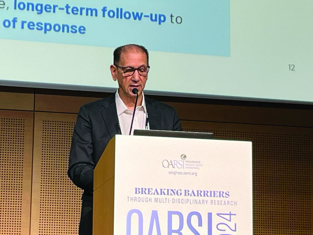
In the same session, James A. Helliwell, MD, cofounder, director, and chief executive officer of Eupraxia Pharmaceuticals in Victoria, British Columbia, Canada, presented updated data from the SPRINGBOARD phase 2B trial of EP-104IAR, a novel long-acting formulation of the corticosteroid fluticasone propionate.
Dr. Helliwell, a cardiothoracic anesthesiologist, explained that EP-104IAR uses proprietary technology to form fluticasone into a crystal that can then be injected directly into the joint. This then slowly diffuses out to provide a highly localized treatment.
The SPRINGBOARD trial recruited just over 300 individuals with moderate knee OA and moderate to severe WOMAC pain and randomly allocated 164 to a single IA injection of EP-104IAR and 164 to a matching vehicle injection as a placebo. The latter was a slightly viscous substance that behaved like hyaluronic acid, Dr. Helliwell said.
The LSM change in total WOMAC score from baseline to week 12 showed a greater improvement with EP-104IAR than with placebo in a per protocol analysis (−2.79 vs −2.07; P = .002). Similar results were seen for the WOMAC subscales of pain (−2.97 vs −2.24; P = .003), function (−2.64 vs −1.99; P = .005), and stiffness (−2.85 vs −2.05; P = .001).
These differences persisted, Dr. Helliwell reported, out to a 20-week assessment for total WOMAC score, function, and stiffness and out to a 15-week assessment for WOMAC pain.
It’s probably no surprise that a steroid works, Dr. Helliwell said, noting that the safety profile of EP-104IAR may be better than that of regular IA steroid injection because it has “few off-target” effects. He reported that there were “minimal, clinically insignificant, and transient effects” of EP-104IAR on serum cortisol. There was no effect on glucose metabolism, even in patients with diabetes, he said.
“There is a group of our patients that we give long-acting steroids to in the joint, so it looked like [the EP-104IAR] safety profile was really good,” Dr. Lane told this news organization. However, she added: “I’m worried about the price tag associated with it.”
PPS
Although it perhaps can’t be described as a novel injectable per se, Mukesh Ahuja, MBBS, global clinical head of osteoarthritis at Paradigm Biopharmaceuticals, presented results of the novel use of PPS.
“PPS is a non-opioid, semi-synthetic xylose-based polysaccharide that is derived from beechwood trees,” Dr. Ahuja said. “It has a long-track record for treating pain, inflammation, and thrombosis in humans.”

There are currently two approved formulations: Oral capsules used for the treatment of interstitial cystitis in the European Union, United States, and Australia and an injectable form used in Italy for thromboprophylaxis.
Dr. Ahuja presented data from a phase 2 trial that looked at the effect of once- or twice-weekly subcutaneous injections of PPS vs placebo in 61 people with knee OA pain. Assessments were made after 56, 168, and 365 days of treatment.
Results showed PPS injections resulted in significant improvements in total WOMAC score, WOMAC pain, and WOMAC function, with more PPS- than placebo-treated individuals achieving and then maintaining at least a 30% or greater improvement in pain and a 56% improvement in function.
Rescue medication use was lower in the PPS-treated patients, and Patient Global Impression of Change were significantly higher, Dr. Ahuja said.
Exploratory analyses of synovial fluid biomarkers showed PPS could be having a direct inflammatory effect, with reductions in several proinflammatory cytokines, such as IL-6 and tumor necrosis factor alpha.
An assessment of OA disease progression using MRI analysis suggested that there may be an effect on cartilage thickness and volume, as well as bone marrow lesions and overall joint inflammation.
Gene Therapy
Elsewhere at OARSI 2024, updated data were reported on XT-150, a non-viral, plasmid-based gene therapy designed to express a proprietary variant (v) of IL-10.
Howard Rutman, MD, MBA, chief medical officer of Xalud Therapeutics, reported data from a patient subgroup analysis of a phase 2 trial, which evaluated the effects of single and repeat IA injections of XT-150.
Previously, it was found that a single dose of XT-150 (0.15 mg/mL or 0.45 mg/mL) given as a 1-mL IA injection did not meet its primary endpoint of a greater proportion of patients achieving a 30% or more improvement in WOMAC pain at 180 days vs a matching placebo.
However, it was noted that 17% of the patients in the trial had a baseline WOMAC pain score of less than 8, so the new analysis focused on a modified intention-to-treat population of 210 patients who had baseline WOMAC pain scores of 9 or higher.
Two injections of XT-150 at a dose of 0.45 mg were found to produce the best effect on WOMAC pain, with a LSM change from baseline of −4.09 vs −2.74 for a single 0.45-mg injection (P = .044).
Dr. Rutman reported that the 0.45-mg dose would be the one moving forward into future studies as this had the best effect when they looked at various patient demographics, including baseline age, gender, body mass index, Kellgren-Lawrence grade, and use of concomitant medications.
XT-150 acts locally, does not integrate into the host genome, and “has a very favorable safety profile,” Dr. Rutman said. As it is not a protein, there is no antibody response, and this gives it the possibility for repeat dosing, with no drug-drug serious adverse events so far reported.
The Best Is Yet to Come?
“There’s a lot of things cooking that haven’t been presented here [at OARSI],” Dr. Lane observed.
“We are figuring out how to regenerate cartilage, and it’s a little different than throwing some stem cells in there. There’s some ground-breaking stuff [coming], it just takes us a while.”
Dr. Lane also noted that researchers were “really figuring out” how joints become painful, which will be a major step in figuring out how to make them less painful for patients.
“We’re making a lot of progress in ways that I don’t think we previously thought of, for example, the weight loss drugs. They probably have a central pain reduction effect, I think there’s a little overlap with the opioid receptors, so that’s pretty exciting. So, we’re getting there,” Dr. Lane said.
The congress was sponsored by the Osteoarthritis Research Society International.
Dr. Lane had no relevant conflicts to declare. The trial of PTP-001 (MOTYS) was funded by Bioventus. Ms. Pavesio is an employee of Doron Therapeutics, a subsidiary of Bioventus. The SPRINGBOARD trial with EP-104IAR was funded by Eupraxia Pharmaceuticals. Dr. Helliwell is an employee and stockholder of Eupraxia Pharmaceuticals. The trial of PPS was funded by Paradigm Biopharmaceuticals. Dr. Ahuja is an employee and stockholder of Paradigm Biopharmaceuticals and holds stock in ChitogenX. The trial of XT-150 was funded by Xalud Therapeutics. Dr. Rutman is an employee and equity holder of the company.
A version of this article appeared on Medscape.com.
VIENNA — Encouraging primary or secondary analyses of trial data for the use of several novel injectables and gene therapy for knee osteoarthritis (OA) were reported at the OARSI 2024 World Congress.
Of all the approaches discussed during the News in Therapies session at OARSI 2024, the most intriguing was the use of the placental extract PTP-001 (MOTYS, Bioventus), session chair Nancy E. Lane, MD, of the University of California Davis School of Medicine, Sacramento, California, told this news organization.
Other notable presentations of data from trials of investigational agents for knee OA included an update from the SPRINGBOARD phase 2B trial of EP-104IAR, a novel long-acting formulation of the corticosteroid fluticasone propionate; a phase 2 trial of pentosan polysulfate sodium (PPS), a non-opioid, semi-synthetic xylose-based polysaccharide; and an update on phase 2 study results for XT-150, a non-viral, plasmid-based gene therapy designed to express a proprietary variant of interleukin 10 (IL-10).
PTP-001 (MOTYS)
Indeed, promising results were seen in a phase 2 trial testing a single intra-articular (IA) injection of PTP-001 vs an IA saline placebo in just over 200 individuals with symptomatic knee OA. Results of this dose-finding study were presented by Alessandra Pavesio, senior vice president and the chief science officer of Bioventus/Doron Therapeutics, Durham, North Carolina.
Ms. Pavesio reported there were decreases in knee pain and improvements in knee function, as measured using the Western Ontario and McMaster Universities Arthritis Index (WOMAC). These changes were seen after 26 weeks of treatment with PTP-001 given at either a low (100 mg, n = 74) or high (200 mg, n = 40) dose.
Although the changes were only numerically and not statistically different from placebo (n = 71) when looking at the total study population, Ms. Pavesio noted that a key objective of the trial had been to identify populations of patients that may benefit.
When they looked at the effects of PTP-001 solely in those with unilateral knee OA, WOMAC pain scores were decreased to a significantly greater extent with both the high and low doses of PTP-001 vs placebo. Decreases in the least squares mean (LSM) change in WOMAC pain from baseline to week 26 were 26.8 with 100-mg PTP-001, 36.1 with 200-mg PTP-001, and 24.0 with placebo (P = .072). A similarly greater effect for PTP-001 was also seen for LSM change in WOMAC function (26.4, 36.0, and 20.0, respectively; P = .023).
Ms. Pavesio noted that the only real side effect seen during the trial was an initial inflammatory reaction within the first 2 days of IA injection, which resolved within a few days without further problems.
The results are promising enough for Ms. Pavesio and her team to consider a phase 3 trial.
Dr. Lane asked Ms. Pavesio: “So, what’s in the secret sauce? You said it was ground-up placentas?” To which Ms. Pavesio replied that it contained about 300 different molecules which came from amnion, chorion, and umbilical cord tissue obtained from consented placental donation.
Dr. Lane subsequently told this news organization: “It’s probably a bunch of growth factors and cytokines, but if it’s not toxic, and they can standardize it, then it might be good. We remain open minded because we haven’t figured it out.”
Novel Fluticasone Delivery
In the same session, James A. Helliwell, MD, cofounder, director, and chief executive officer of Eupraxia Pharmaceuticals in Victoria, British Columbia, Canada, presented updated data from the SPRINGBOARD phase 2B trial of EP-104IAR, a novel long-acting formulation of the corticosteroid fluticasone propionate.
Dr. Helliwell, a cardiothoracic anesthesiologist, explained that EP-104IAR uses proprietary technology to form fluticasone into a crystal that can then be injected directly into the joint. This then slowly diffuses out to provide a highly localized treatment.
The SPRINGBOARD trial recruited just over 300 individuals with moderate knee OA and moderate to severe WOMAC pain and randomly allocated 164 to a single IA injection of EP-104IAR and 164 to a matching vehicle injection as a placebo. The latter was a slightly viscous substance that behaved like hyaluronic acid, Dr. Helliwell said.
The LSM change in total WOMAC score from baseline to week 12 showed a greater improvement with EP-104IAR than with placebo in a per protocol analysis (−2.79 vs −2.07; P = .002). Similar results were seen for the WOMAC subscales of pain (−2.97 vs −2.24; P = .003), function (−2.64 vs −1.99; P = .005), and stiffness (−2.85 vs −2.05; P = .001).
These differences persisted, Dr. Helliwell reported, out to a 20-week assessment for total WOMAC score, function, and stiffness and out to a 15-week assessment for WOMAC pain.
It’s probably no surprise that a steroid works, Dr. Helliwell said, noting that the safety profile of EP-104IAR may be better than that of regular IA steroid injection because it has “few off-target” effects. He reported that there were “minimal, clinically insignificant, and transient effects” of EP-104IAR on serum cortisol. There was no effect on glucose metabolism, even in patients with diabetes, he said.
“There is a group of our patients that we give long-acting steroids to in the joint, so it looked like [the EP-104IAR] safety profile was really good,” Dr. Lane told this news organization. However, she added: “I’m worried about the price tag associated with it.”
PPS
Although it perhaps can’t be described as a novel injectable per se, Mukesh Ahuja, MBBS, global clinical head of osteoarthritis at Paradigm Biopharmaceuticals, presented results of the novel use of PPS.
“PPS is a non-opioid, semi-synthetic xylose-based polysaccharide that is derived from beechwood trees,” Dr. Ahuja said. “It has a long-track record for treating pain, inflammation, and thrombosis in humans.”
There are currently two approved formulations: Oral capsules used for the treatment of interstitial cystitis in the European Union, United States, and Australia and an injectable form used in Italy for thromboprophylaxis.
Dr. Ahuja presented data from a phase 2 trial that looked at the effect of once- or twice-weekly subcutaneous injections of PPS vs placebo in 61 people with knee OA pain. Assessments were made after 56, 168, and 365 days of treatment.
Results showed PPS injections resulted in significant improvements in total WOMAC score, WOMAC pain, and WOMAC function, with more PPS- than placebo-treated individuals achieving and then maintaining at least a 30% or greater improvement in pain and a 56% improvement in function.
Rescue medication use was lower in the PPS-treated patients, and Patient Global Impression of Change were significantly higher, Dr. Ahuja said.
Exploratory analyses of synovial fluid biomarkers showed PPS could be having a direct inflammatory effect, with reductions in several proinflammatory cytokines, such as IL-6 and tumor necrosis factor alpha.
An assessment of OA disease progression using MRI analysis suggested that there may be an effect on cartilage thickness and volume, as well as bone marrow lesions and overall joint inflammation.
Gene Therapy
Elsewhere at OARSI 2024, updated data were reported on XT-150, a non-viral, plasmid-based gene therapy designed to express a proprietary variant (v) of IL-10.
Howard Rutman, MD, MBA, chief medical officer of Xalud Therapeutics, reported data from a patient subgroup analysis of a phase 2 trial, which evaluated the effects of single and repeat IA injections of XT-150.
Previously, it was found that a single dose of XT-150 (0.15 mg/mL or 0.45 mg/mL) given as a 1-mL IA injection did not meet its primary endpoint of a greater proportion of patients achieving a 30% or more improvement in WOMAC pain at 180 days vs a matching placebo.
However, it was noted that 17% of the patients in the trial had a baseline WOMAC pain score of less than 8, so the new analysis focused on a modified intention-to-treat population of 210 patients who had baseline WOMAC pain scores of 9 or higher.
Two injections of XT-150 at a dose of 0.45 mg were found to produce the best effect on WOMAC pain, with a LSM change from baseline of −4.09 vs −2.74 for a single 0.45-mg injection (P = .044).
Dr. Rutman reported that the 0.45-mg dose would be the one moving forward into future studies as this had the best effect when they looked at various patient demographics, including baseline age, gender, body mass index, Kellgren-Lawrence grade, and use of concomitant medications.
XT-150 acts locally, does not integrate into the host genome, and “has a very favorable safety profile,” Dr. Rutman said. As it is not a protein, there is no antibody response, and this gives it the possibility for repeat dosing, with no drug-drug serious adverse events so far reported.
The Best Is Yet to Come?
“There’s a lot of things cooking that haven’t been presented here [at OARSI],” Dr. Lane observed.
“We are figuring out how to regenerate cartilage, and it’s a little different than throwing some stem cells in there. There’s some ground-breaking stuff [coming], it just takes us a while.”
Dr. Lane also noted that researchers were “really figuring out” how joints become painful, which will be a major step in figuring out how to make them less painful for patients.
“We’re making a lot of progress in ways that I don’t think we previously thought of, for example, the weight loss drugs. They probably have a central pain reduction effect, I think there’s a little overlap with the opioid receptors, so that’s pretty exciting. So, we’re getting there,” Dr. Lane said.
The congress was sponsored by the Osteoarthritis Research Society International.
Dr. Lane had no relevant conflicts to declare. The trial of PTP-001 (MOTYS) was funded by Bioventus. Ms. Pavesio is an employee of Doron Therapeutics, a subsidiary of Bioventus. The SPRINGBOARD trial with EP-104IAR was funded by Eupraxia Pharmaceuticals. Dr. Helliwell is an employee and stockholder of Eupraxia Pharmaceuticals. The trial of PPS was funded by Paradigm Biopharmaceuticals. Dr. Ahuja is an employee and stockholder of Paradigm Biopharmaceuticals and holds stock in ChitogenX. The trial of XT-150 was funded by Xalud Therapeutics. Dr. Rutman is an employee and equity holder of the company.
A version of this article appeared on Medscape.com.
FROM OARSI 2024
Do People With Diabetes Need to Fast Longer Before Surgery?
People with diabetes don’t have higher gastric volumes than those without diabetes after following standard preoperative fasting instructions, suggested a study from a team of anesthesiologist researchers.
Moreover, the issue is now further complicated by the widespread use of glucagon-like peptide-1 (GLP-1) receptor agonists for the treatment of both type 2 diabetes and weight loss. These drugs, which were introduced after the study’s enrollment period, work in part by delaying gastric emptying.
The new data come from a prospective study of 84 people with diabetes (85% with type 2) and 96 without diabetes, all with a body mass index (BMI) < 40, who were undergoing elective surgery. A gastric ultrasound was used to assess their gastric contents after they had followed the standard preoperative fasting guidelines of stopping solids 8 hours prior to the procedure and clearing liquids 2 hours prior.
There was no significant difference between the two groups in gastric volume (0.81 mL/kg with diabetes vs 0.87 mL/kg without) or in the proportion with “full stomach,” as designated by the American Society of Anesthesiologists (ASA) guidelines (any solid content or > 1.5 mL/kg of clear fluid), which was seen in 13 with diabetes (15.5%) and 11 (11.5%) without.
Published in Anesthesiology, the findings offer reassurance that different fasting instructions generally aren’t needed for people with diabetes in order to minimize the risk for perioperative pulmonary aspiration, lead author Anahi Perlas, MD, professor of anesthesiology and pain medicine at the University of Toronto, told this news organization.
“We never change practice completely based on a single study, but I think in general, based on our findings, that most diabetic patients aren’t any different from nondiabetics when it comes to their gastric content after fasting, and our standard fasting instructions seem to be just as effective in ensuring an empty stomach.”
But, she added, “If someone has symptoms of gastroparesis or when in doubt, we can always do a gastric ultrasound exam at the bedside and see whether the stomach is full or empty ... it’s very quick, and it’s not difficult to do.”
Expert Identifies Noteworthy Study Limitations
In an accompanying editorial, Mark A. Warner, MD, professor of anesthesiology at the Mayo Clinic in Rochester, Minnesota, said the findings “will be very helpful to anesthesiologists,” although he noted that the exclusion of people with a BMI > 40 is a limitation.
However, Michael Horowitz, MBBS, PhD, FRACP, director of the Endocrine and Metabolic Unit at the Royal Adelaide Hospital and professor of medicine at Adelaide Medical School in Adelaide, Australia, disputed the study’s conclusions. He noted that the sample was small, and the participants had an average A1c of 7.2%. Fewer than half had microvascular or neuropathic complications. Thus, they were healthier than the general population with diabetes.
“They’ve picked the wrong group of diabetics,” said Dr. Horowitz, who specializes in gastrointestinal complications of diabetes. “This is not a group where you would expect a very high prevalence of delayed emptying.”
Gastric emptying of solids and liquids varies widely even among healthy people and more so in those with type 2 diabetes. About a third of those with above-target A1c levels have gastroparesis, while those more in the target range tend to have accelerated emptying, he explained.
And regarding the use of gastric ultrasound for those who are symptomatic, Dr. Horowitz said, “The relationship of symptoms such as nausea, vomiting, fullness, whatever it may be, with the rate of gastric emptying is weak at best. The association is not simply cause and effect.”
Are the Fasting Guidelines Flawed, Regardless of Diabetes Status?
Dr. Horowitz also faulted the ASA’s 2017 guidance revision for allowing clear liquids to be consumed up to 2 hours in advance of anesthesia because it doesn’t distinguish between liquids with and without calories.
“Whether you have diabetes or not, if you are allowed to have a sugar drink up to 2 hours before your operation, the majority of people empty at about 4 kcal/min, so they will still have some of that drink in their stomach,” he said. “If you want an empty stomach, the ASA guidelines are wrong.”
That explains why the study found relatively high rates of “full stomach” in both groups, 15.5% of those with diabetes and 11.5% of those without, he said.
The GLP-1 Agonist Factor
Although the study didn’t address GLP-1 receptor agonist use, Dr. Warner did in his accompanying editorial, noting that the drugs’ rapid expansion “will likely change how we use perioperative fasting guidelines. With these medications delaying gastric emptying times, we now have another risk factor for pulmonary aspiration to consider when applying fasting guidelines. The inconsistent impact of GLP-1 agonists on gastric emptying, ranging from little to significant, makes it difficult for anesthesiologists to gauge whether or not patients taking GLP-1 agonists are likely to have preoperative gastric liquid or solid contents that could cause subsequent damage if regurgitated.”
Gastric ultrasound can be helpful in this situation, Dr. Warner wrote. In addition, he endorsed the 2023 ASA guidance, which calls for withholding daily-dosed GLP-1 agonists on the day of the surgery and the weekly formulations for a week. And if gastrointestinal symptoms are present, delay elective procedures.
But Dr. Horowitz said those recommendations are likely insufficient as well, pointing to data suggesting that daily liraglutide can delay gastric emptying for up to 16 weeks in about a third of patients. Such studies haven’t been conducted by the manufacturers, particularly on the once-weekly formulations, and the ensuing risk for aspiration isn’t known.
“The slowing occurs in much lower doses than are used for glucose lowering,” Dr. Horowitz said. “It is very likely that plasma levels will need to be extremely low to avoid gastric slowing. The current guidelines fail to appreciate this. So, to withhold the short-acting drugs for 1 day is probably wrong. And to stop long-acting drugs for 1 week is almost certainly wrong too.”
But as for what should be done, he said, “I don’t actually know what you do about it. And no one does because there are no data available to answer the question.”
The study received funding from the Physicians’ Services Incorporated Foundation and the Canadian Society of Anesthesiologists. Dr. Perlas received support for nonclinical time through a merit award from the Department of Anesthesiology and Pain Medicine, University of Toronto, and the Department of Anesthesia and Pain Management, Toronto Western Hospital, University Health Network. She is an executive editor of the journal Regional Anesthesia and Pain Medicine and does consulting work for FujiFilm SonoSite. Dr. Horowitz had no relevant disclosures.
A version of this article appeared on Medscape.com.
High-Quality Diet in Early Life May Ward Off Later IBD
, prospective pooled data from two Scandinavian birth cohorts suggested.
It appears important to feed children a quality diet at a very young age, in particular one rich in vegetables and fish, since by age three, only dietary fish intake had any impact on IBD risk.

Although high intakes of these two food categories in very early life correlated with lower IBD risk, exposure to sugar-sweetened beverages (SSBs) was associated with an increased risk. “While non-causal explanations for our results cannot be ruled out, these novel findings are consistent with the hypothesis that early-life diet, possibly mediated through changes in the gut microbiome, may affect the risk of developing IBD,” wrote lead author Annie Guo, a PhD candidate in the Department of Pediatrics, University of Gothenburg, Sweden, and colleagues. The report was published in Gut.
“This is a population-based study investigating the risk for IBD, rather than the specific effect of diet,” Ms. Guo said in an interview. “Therefore, the results are not enough on their own to be translated into individual advice that can be applicable in the clinic. However, the study supports current dietary guidelines for small children, that is, the intake of sugar should be limited and a higher intake of fish and vegetables is beneficial for overall health.”
Two-Cohort Study
The investigators prospectively recorded food-group information on children (just under half were female) from the All Babies in Southeast Sweden and The Norwegian Mother, Father and Child Cohort Study to assess the diet quality using a Healthy Eating Index and intake frequency. Parents answered questions about their offspring’s diet at ages 12-18 months and 30-36 months. Quality of diet was measured by intake of meat, fish, fruit, vegetables, dairy, sweets, snacks, and drinks.
The Swedish cohort included 21,700 children born between October 1997 and October 1999, while the Norwegian analysis included 114,500 children, 95,200 mothers, and 75,200 fathers recruited from across Norway from 1999 to 2008. In 1,304,433 person-years of follow-up, the researchers tracked 81,280 participants from birth to childhood and adolescence, with median follow-ups in the two cohorts ranging from 1 year of age to 21.3 years (Sweden) and to 15.2 years of age (Norway). Of these children, 307 were diagnosed with IBD: Crohn’s disease (CD; n = 131); ulcerative colitis (UC; n = 97); and IBD unclassified (n = 79).
Adjusting for parental IBD history, sex, origin, education, and maternal comorbidities, the study found:
- Compared with low-quality diet, both medium- and high-quality diets at 1 year were associated with a roughly 25% reduced risk for IBD (pooled adjusted hazard ratio [aHR], 0.75 [95% CI, 0.58-0.98] and 0.75 [0.56-1.0], respectively).
- The pooled aHR per increase of category was 0.86 (95% CI, 0.74-0.99). The pooled aHR for IBD in 1-year-olds with high vs low fish intake was 0.70 (95% CI, 0.49-1.0), and this diet showed an association with a reduced risk for UC (pooled aHR, 0.46; 95% CI, 0.21-0.99). Higher vegetable intake at 1 year was also associated with a risk reduction in IBD (HR, 0.72; 95% CI, 0.55-0.95). It has been hypothesized that intake of vegetables and vegetable fibers may have programming effects on the immune system.
- AutoWith 72% of children reportedly consuming SSBs at age 1, pooled aHRs showed that some vs no intake of SSBs was associated with an increased risk for later IBD (pooled aHR, 1.42; 95% CI, 1.05-1.90).
- There were no obvious associations between overall IBD or CD/UC risk and meat, dairy, fruit, grains, potatoes, and foods high in sugar and/or fat. Diet at age 3 years was not associated with incident IBD (pooled aHR, 1.02; 95% CI, 0.76-1.37), suggesting that the risk impact of diet is greatest on very young and vulnerable microbiomes.
Ms. Guo noted that a Swedish national survey among 4-year-olds found a mean SSB consumption of 187 g/d with a mean frequency of once daily. The most desired changes in food habits are a lower intake of soft drinks, sweets, crisps, cakes, and biscuits and an increase in the intake of fruits and vegetables. A similar Norwegian survey among 2-year-olds showed that SSBs were consumed by 36% of all children with a mean intake of 40 g/d.
The exact mechanism by which sugar affects the intestinal microbiota is not established. “However, what we do know is that an excessive intake of sugar can disrupt the balance of the gut microbiome,” Ms. Guo said. “And if the child has a high intake of foods with high in sugar, that also increases the chances that the child’s overall diet has a lower intake of other foods that contribute to a diverse microbiome such as fruits and vegetables.”
An ‘Elegant’ Study
In an accompanying editorial, gastroenterologist Ashwin N. Ananthakrishnan, MBBS, MPH, AGAF, of Mass General Brigham and the Mass General Research Institute, Boston, cautioned that accurately measuring food intake in very young children is difficult, and dietary questionnaires in this study did not address food additives and emulsifiers common in commercial baby food, which may play a role in the pathogenesis of IBD.

Another study limitation is that the dietary questionnaire used has not been qualitatively or quantitatively validated against other more conventional methods, said Dr. Ananthakrishnan, who was not involved in the research.
Nevertheless, he called the study “elegant” and expanding of the data on the importance of this period in IBD development. “Although in the present study there was no association between diet at 3 years and development of IBD (in contrast to the association observed for dietary intake at 1 year), other prospective cohorts of adult-onset IBD have demonstrated an inverse association between vegetable or fish intake and reduced risk for CD while sugar-sweetened beverages have been linked to a higher risk for IBD.”
As to the question of recommending early preventive diet for IBD, “thus far, data on the impact of diet very early in childhood, outside of breastfeeding, on the risk for IBD has been lacking,” Dr. Ananthakrishnan said in an interview. “This important study highlights that diet as early as 1 year can modify subsequent risk for IBD. This raises the intriguing possibility of whether early changes in diet could be used, particularly in those at higher risk, to reduce or even prevent future development of IBD. Of course, more works needs to be done to define modifiability of diet as a risk factor, but this is an important supportive data.”
In his editorial, Dr. Ananthakrishnan stated that despite the absence of gold-standard interventional data demonstrating a benefit of dietary interventions, “in my opinion, it may still be reasonable to suggest such interventions to motivate individuals who incorporate several of the dietary patterns associated with lower risk for IBD from this and other studies. This includes ensuring adequate dietary fiber, particularly from fruits and vegetables, intake of fish, minimizing sugar-sweetened beverages and preferring fresh over processed and ultra-processed foods and snacks.” According to the study authors, their novel findings support further research on the role of childhood diet in the prevention of IBD.
The All Babies in Southeast Sweden Study is supported by Barndiabetesfonden (Swedish Child Diabetes Foundation), the Swedish Council for Working Life and Social Research, the Swedish Research Council, the Medical Research Council of Southeast Sweden, the JDRF Wallenberg Foundation, ALF and LFoU grants from Region Östergötland and Linköping University, and the Joanna Cocozza Foundation.
The Norwegian Mother, Father and Child Cohort Study is supported by the Norwegian Ministry of Health and Care Services and the Ministry of Education and Research.
Ms. Guo received grants from the Swedish Society for Medical Research and the Henning and Johan Throne-Holst Foundation to conduct this study. Co-author Karl Mårild has received funding from the Swedish Society for Medical Research, the Swedish Research Council, and ALF, Sweden’s medical research and education co-ordinating body. The authors declared no competing interests. Dr. Ananthakrishnan is supported by the National Institutes of Health, the Leona M. and Harry B. Helmsley Charitable Trust, and the Chleck Family Foundation. He has served on the scientific advisory board for Geneoscopy.
, prospective pooled data from two Scandinavian birth cohorts suggested.
It appears important to feed children a quality diet at a very young age, in particular one rich in vegetables and fish, since by age three, only dietary fish intake had any impact on IBD risk.
Although high intakes of these two food categories in very early life correlated with lower IBD risk, exposure to sugar-sweetened beverages (SSBs) was associated with an increased risk. “While non-causal explanations for our results cannot be ruled out, these novel findings are consistent with the hypothesis that early-life diet, possibly mediated through changes in the gut microbiome, may affect the risk of developing IBD,” wrote lead author Annie Guo, a PhD candidate in the Department of Pediatrics, University of Gothenburg, Sweden, and colleagues. The report was published in Gut.
“This is a population-based study investigating the risk for IBD, rather than the specific effect of diet,” Ms. Guo said in an interview. “Therefore, the results are not enough on their own to be translated into individual advice that can be applicable in the clinic. However, the study supports current dietary guidelines for small children, that is, the intake of sugar should be limited and a higher intake of fish and vegetables is beneficial for overall health.”
Two-Cohort Study
The investigators prospectively recorded food-group information on children (just under half were female) from the All Babies in Southeast Sweden and The Norwegian Mother, Father and Child Cohort Study to assess the diet quality using a Healthy Eating Index and intake frequency. Parents answered questions about their offspring’s diet at ages 12-18 months and 30-36 months. Quality of diet was measured by intake of meat, fish, fruit, vegetables, dairy, sweets, snacks, and drinks.
The Swedish cohort included 21,700 children born between October 1997 and October 1999, while the Norwegian analysis included 114,500 children, 95,200 mothers, and 75,200 fathers recruited from across Norway from 1999 to 2008. In 1,304,433 person-years of follow-up, the researchers tracked 81,280 participants from birth to childhood and adolescence, with median follow-ups in the two cohorts ranging from 1 year of age to 21.3 years (Sweden) and to 15.2 years of age (Norway). Of these children, 307 were diagnosed with IBD: Crohn’s disease (CD; n = 131); ulcerative colitis (UC; n = 97); and IBD unclassified (n = 79).
Adjusting for parental IBD history, sex, origin, education, and maternal comorbidities, the study found:
- Compared with low-quality diet, both medium- and high-quality diets at 1 year were associated with a roughly 25% reduced risk for IBD (pooled adjusted hazard ratio [aHR], 0.75 [95% CI, 0.58-0.98] and 0.75 [0.56-1.0], respectively).
- The pooled aHR per increase of category was 0.86 (95% CI, 0.74-0.99). The pooled aHR for IBD in 1-year-olds with high vs low fish intake was 0.70 (95% CI, 0.49-1.0), and this diet showed an association with a reduced risk for UC (pooled aHR, 0.46; 95% CI, 0.21-0.99). Higher vegetable intake at 1 year was also associated with a risk reduction in IBD (HR, 0.72; 95% CI, 0.55-0.95). It has been hypothesized that intake of vegetables and vegetable fibers may have programming effects on the immune system.
- AutoWith 72% of children reportedly consuming SSBs at age 1, pooled aHRs showed that some vs no intake of SSBs was associated with an increased risk for later IBD (pooled aHR, 1.42; 95% CI, 1.05-1.90).
- There were no obvious associations between overall IBD or CD/UC risk and meat, dairy, fruit, grains, potatoes, and foods high in sugar and/or fat. Diet at age 3 years was not associated with incident IBD (pooled aHR, 1.02; 95% CI, 0.76-1.37), suggesting that the risk impact of diet is greatest on very young and vulnerable microbiomes.
Ms. Guo noted that a Swedish national survey among 4-year-olds found a mean SSB consumption of 187 g/d with a mean frequency of once daily. The most desired changes in food habits are a lower intake of soft drinks, sweets, crisps, cakes, and biscuits and an increase in the intake of fruits and vegetables. A similar Norwegian survey among 2-year-olds showed that SSBs were consumed by 36% of all children with a mean intake of 40 g/d.
The exact mechanism by which sugar affects the intestinal microbiota is not established. “However, what we do know is that an excessive intake of sugar can disrupt the balance of the gut microbiome,” Ms. Guo said. “And if the child has a high intake of foods with high in sugar, that also increases the chances that the child’s overall diet has a lower intake of other foods that contribute to a diverse microbiome such as fruits and vegetables.”
An ‘Elegant’ Study
In an accompanying editorial, gastroenterologist Ashwin N. Ananthakrishnan, MBBS, MPH, AGAF, of Mass General Brigham and the Mass General Research Institute, Boston, cautioned that accurately measuring food intake in very young children is difficult, and dietary questionnaires in this study did not address food additives and emulsifiers common in commercial baby food, which may play a role in the pathogenesis of IBD.
Another study limitation is that the dietary questionnaire used has not been qualitatively or quantitatively validated against other more conventional methods, said Dr. Ananthakrishnan, who was not involved in the research.
Nevertheless, he called the study “elegant” and expanding of the data on the importance of this period in IBD development. “Although in the present study there was no association between diet at 3 years and development of IBD (in contrast to the association observed for dietary intake at 1 year), other prospective cohorts of adult-onset IBD have demonstrated an inverse association between vegetable or fish intake and reduced risk for CD while sugar-sweetened beverages have been linked to a higher risk for IBD.”
As to the question of recommending early preventive diet for IBD, “thus far, data on the impact of diet very early in childhood, outside of breastfeeding, on the risk for IBD has been lacking,” Dr. Ananthakrishnan said in an interview. “This important study highlights that diet as early as 1 year can modify subsequent risk for IBD. This raises the intriguing possibility of whether early changes in diet could be used, particularly in those at higher risk, to reduce or even prevent future development of IBD. Of course, more works needs to be done to define modifiability of diet as a risk factor, but this is an important supportive data.”
In his editorial, Dr. Ananthakrishnan stated that despite the absence of gold-standard interventional data demonstrating a benefit of dietary interventions, “in my opinion, it may still be reasonable to suggest such interventions to motivate individuals who incorporate several of the dietary patterns associated with lower risk for IBD from this and other studies. This includes ensuring adequate dietary fiber, particularly from fruits and vegetables, intake of fish, minimizing sugar-sweetened beverages and preferring fresh over processed and ultra-processed foods and snacks.” According to the study authors, their novel findings support further research on the role of childhood diet in the prevention of IBD.
The All Babies in Southeast Sweden Study is supported by Barndiabetesfonden (Swedish Child Diabetes Foundation), the Swedish Council for Working Life and Social Research, the Swedish Research Council, the Medical Research Council of Southeast Sweden, the JDRF Wallenberg Foundation, ALF and LFoU grants from Region Östergötland and Linköping University, and the Joanna Cocozza Foundation.
The Norwegian Mother, Father and Child Cohort Study is supported by the Norwegian Ministry of Health and Care Services and the Ministry of Education and Research.
Ms. Guo received grants from the Swedish Society for Medical Research and the Henning and Johan Throne-Holst Foundation to conduct this study. Co-author Karl Mårild has received funding from the Swedish Society for Medical Research, the Swedish Research Council, and ALF, Sweden’s medical research and education co-ordinating body. The authors declared no competing interests. Dr. Ananthakrishnan is supported by the National Institutes of Health, the Leona M. and Harry B. Helmsley Charitable Trust, and the Chleck Family Foundation. He has served on the scientific advisory board for Geneoscopy.
FROM GUT
The Inconsistency of Preparticipation Sports Evaluations Raises Issues About Their Utility
TORONTO — There is little consistency in the elements and types of information captured in preparticipation physical evaluations (PPE) for sports among school-aged children, which is complicating efforts to determine if they have value, according to a study presented at the Pediatric Academic Societies annual meeting.
The study concept developed when Tammy Ng, MD, a third-year resident in pediatrics at the University of California, Davis, School of Medicine in Sacramento, was surprised to learn that the American Academy of Pediatrics (AAP) had been issuing a standard-of-care PPE for decades.
Dr. Ng had a long-standing interest in pediatric sports medicine and thought that if she was unfamiliar with this form, which was first developed by the AAP in the 1990s in collaboration with other professional organizations, there must be others who were unaware of this resource.
Assuming that this collaborative effort led by the AAP could serve as a standard of care, Dr. Ng evaluated whether PPEs at her own institution were capturing similar information.
In the most recent (5th) edition of the PPE, which was released in 2019 and is available online, medical history is elicited for numerous organ systems relevant to risk. The questions are not directed to any specific sport; the form does not even provide a question about which sports are being considered.
Little Consistency
In evaluating whether PPEs completed at her institution in the previous year elicited similar information, Dr. Ng sought to match 25 elements of patient history from the AAP form to questions posed in the PPEs completed at her institution, some of which had been supplied by school or sports organizations.
Of the 365 PPE forms completed at Dr. Ng’s institution that met study criteria, only 28.6% addressed all 25 elements in the AAP form (range, 0%-78%). Although more than half asked specifically about a history of respiratory symptoms, fewer than half included inquiries about cardiovascular history. There was also little consistency in the capture of information about other relevant medical history.
According to Dr. Ng, these low percentages were observed even when liberally awarding credit. For one example, she said forms that asked any question about syncope with exercise were credited with seeking information about cardiovascular health even though a yes-or-no response might not be helpful.
“We did not distinguish between syncope before or after exercise and this is relevant,” Dr. Ng said. “Syncope during exercise is more likely to be a predictor of sudden cardiac death, whereas syncope after exercise is more likely to be a vasovagal response to exertion.”
Of the 365 PPEs evaluated, about half were completed by pediatricians and half by family medicine clinicians. The average age of the children was about 14 years. Sixty-three percent were male. Only one third of the forms documented the sport for which a pre-participation screen was being submitted.
While almost all states now require PPEs for children considering participation in sports, few specify what information should be elicited, according to Dr. Ng. She further noted that no major study has shown that PPEs have any role in preventing morbidity or mortality related to sports participation.
Does Heterogeneity Negate Worth?
With such diversity across PPEs, evaluating their role is difficult. For example, with such heterogeneity among forms for the information elicited, there is no reasonable approach for testing their sensitivity in predicting medical complications.
Dr. Ng noted that school-created forms were just as likely as forms from other sources to diverge from the AAP-endorsed PPE and ignore organ systems relevant to risk of medical complications. Yet, if the answer is to use the AAP form, Dr. Ng noted that the first sentence on the form reads, “This form should be placed in the athlete’s medical file and should not be shared with schools or sports organizations.”
Although Dr. Ng acknowledged that providing completed PPEs to third parties raises questions about privacy, she questioned how the information should be used by children, parents, and sports organization administrators for discussing risks if not shared.
This concern was seconded in the discussion following Dr. Ng’s presentation.
“You might be signing off on sports participation, but is this for cheerleading or for football?” asked Daniel C. Worthington, MD, a pediatrician in private practice who has a clinical appointment at Case Western Reserve University School of Medicine, Cleveland. “This makes a huge difference when evaluating if participation is safe.”
He has no issue with completing PPEs for the goal of keeping children safe, but he focused on the inconsistency of how information is collected and distributed.
“The major question is: Does it make any difference?” said Dr. Worthington, referring to the completion of PPEs.
Another participant in the discussion that followed Dr. Ng’s presentation pointed out that the urgent care office in a mall near to his office offers a completed PPE form for a price of $20. In their recommendations, the AAP suggests PPEs be completed by the individual’s primary care physician during a well visit, according to Dr. Ng.
Dr. Ng indicated that PPEs and their purpose deserve a closer look. Based on her data, it is reasonable to assume that the priority for some – whether those requiring or those completing the form — is completing the task rather than meaningful screening of risk.
Dr. Ng and Dr. Worthington report no potential conflicts of interest.
FROM PAS 2024
Neutrophils Take Center Stage in Growing Understanding of Colchicine’s Role in Treating Atherosclerotic Cardiovascular Disease
NEW YORK — New insights into colchicine’s disruption of the pathway that contributes to arterial inflammation and new clinical studies of the drug could pave the way toward greater use of the anti-inflammatory drug in patients with or at risk for atherosclerotic cardiovascular disease (ASCVD), researchers said at the 4th Annual Cardiometabolic Risk in Inflammatory Conditions conference.
Colchicine was approved by the US Food and Drug Administration (FDA) in June 2023 in a once-daily 0.5-mg formulation under the brand name Lodoco to reduce the risk for major adverse cardiovascular events (MACE) in patients with established atherosclerotic disease or with multiple risk factors for CVD. The Lodoco formulation is slightly smaller than the 0.6-mg formulation that’s taken twice daily for the prophylaxis and treatment of acute gout flares.
In a presentation at the conference, Binita Shah, MD, one of the principal investigators in trials of Lodoco, explained how the inflammatory pathway contributes to atherosclerosis and provided an update on how colchicine disrupts the pathway. Dr. Shah is an associate professor of medicine at New York University in New York City and director of research at NYU Langone Health Interventional Cardiology.
“Colchicine dampens inflammatory markers on neutrophils so that they are less likely to be attracted to inflamed or injured endothelium, which would be the site of where plaque is building up or where the plaque has ruptured in the setting of a heart attack,” Shah told this news organization after her presentation.

The Inflammatory Pathway
Dr. Shah explained that normal coronary endothelium resists adhesion by circulating leukocytes, but inflamed or injured coronary endothelium attracts those neutrophils via two types of selectins: L-selectins on neutrophils and E-selectins on endothelial cells. Those neutrophils then release inflammatory cytokines including interleukin-1 beta (IL-1ß), which then triggers production of IL-6 and, subsequently, high-sensitivity C-reactive protein (hsCRP), which contributes to plaque formation, she said.
“Colchicine affects these pathways with a balance for safety and effect on clinical outcomes, particularly to reduce recurrent myocardial infarction [MI],” Dr. Shah said during her presentation.
Results from the CIRT trial demonstrated that methotrexate is ineffective in blocking the adenosine-mediated anti-inflammatory pathway, Dr. Shah said, so focusing on the IL-1ß–IL-6–hsCRP pathway, which is known to work based on the results of the CANTOS trial, could pay dividends.
“This is where colchicine can potentially play a role,” she said.
Dr. Shah cited a secondary analysis of the CANTOS trial in which the magnitude of hsCRP reduction correlated with a reduction in MI, stroke, or cardiovascular death. The secondary analysis showed that patients who received canakinumab and achieved hsCRP ≥ 2 mg/L had a nonsignificant 5% lower risk and those who reached < 2 mg/L had a statistically significant 25% lower risk than those who received placebo.
The COPE-PCI Pilot trial demonstrated the benefit of targeting the interleukin pathways, she noted.
Further clarification of the role of colchicine in managing patients with acute coronary syndrome may come from two other randomized trials now underway, Dr. Shah said: POPCORN is evaluating colchicine to reduce MACE after noncardiac surgery, and CLEAR SYNERGY is evaluating the best timing for colchicine therapy after an acute MI.
Dr. Shah presented preliminary data from her group from a neutrophil biomarker substudy of CLEAR SYNERGY that isolated neutrophils from patients who had an acute MI. “We treated them with various doses of colchicine and showed that the interaction between those treated neutrophils [and] the endothelial cells were a lot lower; they were less sticky to endothelial cells as colchicine was administered,” she said in her presentation. She added that colchicine also reduced neutrophil chemotaxis and neutrophil activation and potentially inhibited inflammasomes, decreasing IL-1ß production.
What’s more, colchicine has been shown to not affect platelets alone but rather platelets at the site of inflammation or plaque rupture, Dr. Shah added. “At currently used doses, colchicine does not inhibit platelet activity [by] itself, so we’ve never seen increased bleeding events, but it will dampen neutrophils’ ability to latch onto a platelet that could contribute to a clot,” she later told this news organization.
“There are multiple studies, both retrospective studies in gout cohorts as well as prospective studies in the cardiovascular cohort, that all show consistently one thing, which is that colchicine continues to reduce the risk of having a recurrent MI in patients who either have cardiovascular disease or are at high risk of having cardiovascular disease,” she said.
“I think that’s very helpful to know that it’s not just one study — it’s not just a fluke, potentially a play of chance — but multiple studies consistently showing the same thing: That there’s a reduced risk of acute MI.”
Slow to Embrace Colchicine
Despite this evidence, cardiologists and rheumatologists have been slow to embrace colchicine for patients at risk for cardiovascular events, said Michael S. Garshick, MD, who attended the conference and is head of the Cardio-Rheumatology Program at NYU Langone. “What [Shah] really highlighted was that for a number of years now, we’ve had several clinical trials showing the benefit of low-dose colchicine to prevent atherosclerotic cardiovascular events, and yet despite these and that there’s now an indication to use low-dose colchicine to reduce cardiovascular disease, we’re still struggling for this medication to be taken up by the general cardiology community to treat high-risk patients.

“There’s still some work to do to prove that we need to break those barriers,” Dr. Garshick added. Some of the confusion surrounding the use of colchicine for ASCVD may be attributed to the 0.5-mg dose approved for CVD as opposed to the long-approved 0.6-mg dose for gout, he said. “People are generally confused: Is it OK to use the 0.6-mg dose?” Dr. Garshick said.
Potential gastrointestinal side effects may be another concerning factor, although, he added, “we didn’t see any major complications.” Another issue could be polypharmacy in many of these patients, he said.
Dr. Garshick concurred with Shah that the existing evidence supporting the use of colchicine to reduce risk for cardiovascular events is strong, but more will come out. “I think there’s going to be evolving data supporting it,” he said.
Dr. Shah disclosed financial relationships with Philips Volcano and Novo Nordisk. She is a principal investigator of the CLEAR SYNERGY biomarker substudy and the POPCORN trial. Dr. Garshick disclosed relationships with Kiniksa Pharmaceuticals, Agepha Pharma, Bristol Myers Squibb, and Horizon Therapeutics.
A version of this article appeared on Medscape.com .
NEW YORK — New insights into colchicine’s disruption of the pathway that contributes to arterial inflammation and new clinical studies of the drug could pave the way toward greater use of the anti-inflammatory drug in patients with or at risk for atherosclerotic cardiovascular disease (ASCVD), researchers said at the 4th Annual Cardiometabolic Risk in Inflammatory Conditions conference.
Colchicine was approved by the US Food and Drug Administration (FDA) in June 2023 in a once-daily 0.5-mg formulation under the brand name Lodoco to reduce the risk for major adverse cardiovascular events (MACE) in patients with established atherosclerotic disease or with multiple risk factors for CVD. The Lodoco formulation is slightly smaller than the 0.6-mg formulation that’s taken twice daily for the prophylaxis and treatment of acute gout flares.
In a presentation at the conference, Binita Shah, MD, one of the principal investigators in trials of Lodoco, explained how the inflammatory pathway contributes to atherosclerosis and provided an update on how colchicine disrupts the pathway. Dr. Shah is an associate professor of medicine at New York University in New York City and director of research at NYU Langone Health Interventional Cardiology.
“Colchicine dampens inflammatory markers on neutrophils so that they are less likely to be attracted to inflamed or injured endothelium, which would be the site of where plaque is building up or where the plaque has ruptured in the setting of a heart attack,” Shah told this news organization after her presentation.
The Inflammatory Pathway
Dr. Shah explained that normal coronary endothelium resists adhesion by circulating leukocytes, but inflamed or injured coronary endothelium attracts those neutrophils via two types of selectins: L-selectins on neutrophils and E-selectins on endothelial cells. Those neutrophils then release inflammatory cytokines including interleukin-1 beta (IL-1ß), which then triggers production of IL-6 and, subsequently, high-sensitivity C-reactive protein (hsCRP), which contributes to plaque formation, she said.
“Colchicine affects these pathways with a balance for safety and effect on clinical outcomes, particularly to reduce recurrent myocardial infarction [MI],” Dr. Shah said during her presentation.
Results from the CIRT trial demonstrated that methotrexate is ineffective in blocking the adenosine-mediated anti-inflammatory pathway, Dr. Shah said, so focusing on the IL-1ß–IL-6–hsCRP pathway, which is known to work based on the results of the CANTOS trial, could pay dividends.
“This is where colchicine can potentially play a role,” she said.
Dr. Shah cited a secondary analysis of the CANTOS trial in which the magnitude of hsCRP reduction correlated with a reduction in MI, stroke, or cardiovascular death. The secondary analysis showed that patients who received canakinumab and achieved hsCRP ≥ 2 mg/L had a nonsignificant 5% lower risk and those who reached < 2 mg/L had a statistically significant 25% lower risk than those who received placebo.
The COPE-PCI Pilot trial demonstrated the benefit of targeting the interleukin pathways, she noted.
Further clarification of the role of colchicine in managing patients with acute coronary syndrome may come from two other randomized trials now underway, Dr. Shah said: POPCORN is evaluating colchicine to reduce MACE after noncardiac surgery, and CLEAR SYNERGY is evaluating the best timing for colchicine therapy after an acute MI.
Dr. Shah presented preliminary data from her group from a neutrophil biomarker substudy of CLEAR SYNERGY that isolated neutrophils from patients who had an acute MI. “We treated them with various doses of colchicine and showed that the interaction between those treated neutrophils [and] the endothelial cells were a lot lower; they were less sticky to endothelial cells as colchicine was administered,” she said in her presentation. She added that colchicine also reduced neutrophil chemotaxis and neutrophil activation and potentially inhibited inflammasomes, decreasing IL-1ß production.
What’s more, colchicine has been shown to not affect platelets alone but rather platelets at the site of inflammation or plaque rupture, Dr. Shah added. “At currently used doses, colchicine does not inhibit platelet activity [by] itself, so we’ve never seen increased bleeding events, but it will dampen neutrophils’ ability to latch onto a platelet that could contribute to a clot,” she later told this news organization.
“There are multiple studies, both retrospective studies in gout cohorts as well as prospective studies in the cardiovascular cohort, that all show consistently one thing, which is that colchicine continues to reduce the risk of having a recurrent MI in patients who either have cardiovascular disease or are at high risk of having cardiovascular disease,” she said.
“I think that’s very helpful to know that it’s not just one study — it’s not just a fluke, potentially a play of chance — but multiple studies consistently showing the same thing: That there’s a reduced risk of acute MI.”
Slow to Embrace Colchicine
Despite this evidence, cardiologists and rheumatologists have been slow to embrace colchicine for patients at risk for cardiovascular events, said Michael S. Garshick, MD, who attended the conference and is head of the Cardio-Rheumatology Program at NYU Langone. “What [Shah] really highlighted was that for a number of years now, we’ve had several clinical trials showing the benefit of low-dose colchicine to prevent atherosclerotic cardiovascular events, and yet despite these and that there’s now an indication to use low-dose colchicine to reduce cardiovascular disease, we’re still struggling for this medication to be taken up by the general cardiology community to treat high-risk patients.
“There’s still some work to do to prove that we need to break those barriers,” Dr. Garshick added. Some of the confusion surrounding the use of colchicine for ASCVD may be attributed to the 0.5-mg dose approved for CVD as opposed to the long-approved 0.6-mg dose for gout, he said. “People are generally confused: Is it OK to use the 0.6-mg dose?” Dr. Garshick said.
Potential gastrointestinal side effects may be another concerning factor, although, he added, “we didn’t see any major complications.” Another issue could be polypharmacy in many of these patients, he said.
Dr. Garshick concurred with Shah that the existing evidence supporting the use of colchicine to reduce risk for cardiovascular events is strong, but more will come out. “I think there’s going to be evolving data supporting it,” he said.
Dr. Shah disclosed financial relationships with Philips Volcano and Novo Nordisk. She is a principal investigator of the CLEAR SYNERGY biomarker substudy and the POPCORN trial. Dr. Garshick disclosed relationships with Kiniksa Pharmaceuticals, Agepha Pharma, Bristol Myers Squibb, and Horizon Therapeutics.
A version of this article appeared on Medscape.com .
Do Health-Related Social Needs Raise Mortality Risk in Cancer Survivors?
Little is known about the specific association between health-related social needs (HRSNs) and mortality risk even though HRSNs, defined as challenges in affording food, housing, and other necessities of daily living, are potential challenges for cancer survivors, wrote Zhiyuan Zheng, PhD, of the American Cancer Society, Atlanta, and colleagues.
A 2020 study by Dr. Zheng and colleagues published in the Journal of the National Comprehensive Cancer Network (NCCN) showed that food insecurity and financial worries had a negative impact on cancer survivorship. In the new study, published in Cancer, the researchers identified cancer survivors using the 2013-2018 National Health Interview Survey (NHIS) and the NHIS Mortality File through December 31, 2019. The researchers examined mortality using the data from the Centers for Disease Control and Prevention’s National Death Index (NDI) through December 31, 2019, which links to the National Health Interview Survey Data used in the study.
Individuals’ HRSNs were categorized into three groups: severe, moderate, and minor/none. HRSNs included food insecurity and nonmedical financial concerns, such as housing costs (rent, mortgage). Medical financial hardship included material, psychological, and behavioral domains and was divided into three groups: 2-3 domains, 1 domain, or 0 domains.
What Are the Potential Financial Implications of this Research?
The high costs of cancer care often cause medical financial hardships for cancer survivors, and expenses also may cause psychological distress and nonmedical financial hardship as survivors try to make ends meet while facing medical bills, wrote Dr. Zheng and colleagues.
Policy makers are increasingly interested in adding HRSNs to insurance coverage; recent guidance from the Centers for Medicare & Medicaid Services (CMS) allows individual states to apply to provide nutrition and housing supports through state Medicaid programs, according to authors of a 2023 article published in JAMA Health Forum.
The new study adds to the understanding of how HRSNs impact people with cancer by examining the association with mortality risk, Yelak Biru, MSc, president and chief executive officer of the International Myeloma Foundation, said in an interview.
“This is a key area of study for addressing the disparities in treatments and outcomes that result in inequities,” said Mr. Biru, a patient advocate and multiple myeloma survivor who was not involved in the study.
What Does the New Study Show?
The new study characterized HRSNs in 5,855 adult cancer survivors aged 18-64 years and 5,918 aged 65-79 years. In the 18- to 64-year-old group, 25.5% reported moderate levels of HRSNs, and 18.3% reported severe HRSNs. In patients aged 65-79 years, 15.6% and 6.6% reported moderate HRSNs and severe HRSNs, respectively.
Severe HRSN was significantly associated with higher mortality risk in an adjusted analysis in patients aged 18-64 years (hazard ratio 2.00, P < .001).
Among adults aged 65-79 years, severe HRSN was not associated with higher mortality risk; however, in this older age group, those with 2-3 domains of medical financial hardship had significantly increased mortality risk compared with adults aged 65-79 years with zero domains of medical financial hardship (HR 1.58, P = .007).
Although the findings that HRSNs were associated with increased mortality risk, especially in the younger group, were not surprising, they serve as a call to action to address how HRSNs are contributing to cancer mortality, Mr. Biru said in an interview. “HRSNs, like food or housing insecurity, can lead to patients being unable to undergo the best treatment approach for their cancer,” he said.
What Are the Limitations and Research Gaps?
The study findings were limited by several factors including the use of self-reports to measure medical financial hardship, food insecurity, and nonmedical financial concerns in the NHIS, the researchers wrote in their discussion. More research with longer follow-up time beyond 1-5 years is needed, wrote Dr. Zheng and colleagues.
Studies also are needed to illustrate how patient navigation can help prevent patients from falling through the cracks with regard to social needs and financial hardships, Mr. Biru told this news organization.
Other areas for research include how addressing social needs affects health outcomes and whether programs designed to address social needs are effective, he said.
“Finally, qualitative research is needed to capture the lived experiences of cancer survivors facing these challenges. This knowledge can inform the development of more patient-centered interventions and policies that effectively address the social determinants of health and improve overall outcomes for all cancer survivors,” Mr. Biru said.
What Is the Takeaway Message for Clinicians?
HRSNs and financial hardship are significantly associated with increased risk of mortality in adult cancer survivors, Dr. Zheng and colleagues concluded. Looking ahead, comprehensive assessment of HRSNs and financial hardship may help clinicians connect patients with relevant services to mitigate the social and financial impacts of cancer, they wrote.
“The takeaway message for oncologists in practice is that addressing [HRSNs] and financial hardship is crucial for providing comprehensive and equitable cancer care,” Mr. Biru said during his interview.
“The impact of social determinants of health on cancer outcomes cannot be ignored, and oncologists play a vital role in identifying and addressing these needs,” he said. Sensitive, discussion-based screenings are needed to identify core needs such as food and transportation, but clinicians also can consider broader social factors and work with a team to connect patients to appropriate resources, he added.
“By recognizing the importance of HRSN screening and taking proactive steps to address these needs, oncologists can contribute to improving health outcomes, reducing healthcare disparities, and providing more equitable cancer care for their patients,” he said.
What Other Guidance Is Available?
“High-quality cancer care requires treating the whole person; measuring and addressing anything in their life that could result in poorer health outcomes is a key component of comprehensive care,” Mr. Biru emphasized.
In September 2023, the National Comprehensive Cancer Network (NCCN) convened a working group cochaired by Mr. Biru that developed recommendations for how oncology practices should routinely measure HRSNs (NCCN.org/social-needs).
“The working group proposed that every cancer patient be assessed for food, transportation access, and financial and housing security at least once a year, and be reassessed at every care transition point as well,” Mr. Biru said. Such screenings should include follow-up to connect patients with services to address any HRSNs they are experiencing, he added.
Lead author Dr. Zheng is employed by the American Cancer Society, which as a nonprofit receives funds from the public through fundraising and contributions, as well as some support from corporations and industry to support its mission programs and services. Mr. Biru had no financial conflicts to disclose.
FROM CANCER
ADHD Tied to Risk for Lewy Body Disease, Dementia, MCI
, results of a new study showed.
“Determining whether there is an association between ADHD and subsequent conversion to a specific type of dementia is important. This information could generate opportunities for prevention and early treatment, as well as initiate research into the pathophysiological processes involved in understanding the process of cognitive decline,” the researchers, led by Ángel Golimstok, MD, of Hospital Italiano, Buenos Aires, Argentina, wrote.
The findings were published online in The American Journal of Geriatric Psychiatry.
Seeking Confirmation
The researchers first identified a link between DLB and ADHD in 2011. Since then, there have been eight additional studies from other groups also showing a possible link between ADHD and DLB.
To confirm the relationship, the researchers recruited 270 individuals between the ages of 45 and 70 years between 2007 and 2012. Of these, 161 had ADHD, and 109 were healthy controls.
Participants with ADHD met the Diagnostic and Statistical Manual of Mental Disorders, Fourth Edition, text revision criteria for a diagnosis in the past and had a chronic course of ADHD symptoms from adolescence to adulthood that caused mild to severe impairment.
Investigators excluded participants who had been taking ADHD medications for 6 months or more, those with MCI at study initiation, and those with other comorbid psychiatric disorders.
At baseline, all participants received a physical exam, an MRI, and a neuropsychological exam to test for any type of dementia-related impairment.
Study participants were followed for an average of 12 years. A total of 27 individuals with ADHD developed dementia versus four patients in the control group (17% vs 4%, respectively), and 19 of those also had DLB (P = .002 for both).
Of those who developed any type of dementia, 87% were from the ADHD group. The most frequent type of dementia was DLB, 95% of which occurred in the ADHD group. Overall, DLB represented 70% of the dementia cases among participants with ADHD.
A total of 108 participants with ADHD were subsequently diagnosed with naMCI versus 19 healthy controls (67% vs 17%; P < .001).
“Although this pattern of deficits is reasonably expected in early DLB, these results should be interpreted with caution because they may be related to the overlap of symptoms and cognitive deficits between ADHD and naMCI, which may lead to an overestimation of the degenerative phenomenon. Thus, our cases of naMCI could correspond to the natural aging of ADHD patients and not to pathological deterioration,” the authors wrote.
The researchers pointed out that the sample of patients with ADHD originally sought evaluation because of a cognitive complaint or their own motivation. Therefore, the study results are not generalizable to all patients with ADHD. Another limitation was the relatively small number of patients included in the sample.
There was no reported source of funding, and there were no relevant disclosures reported.
A version of this article appeared on Medscape.com.
FROM THE AMERICAN JOURNAL OF GERIATRIC PSYCHIATRY
Proton Pump Inhibitors Tied to Migraine, Other Severe Headache Types
, new research showed.
Using data from the National Health and Nutrition Examination Survey (NHANES), investigators conducted a cross-sectional analysis and found all types of acid-suppression therapy were associated with an increased risk for severe headache including migraine but that PPIs conferred the greatest risk.
“It’s important to note that many people do need acid-reducing medications to manage acid reflux or other conditions, and people with migraine or severe headache who are taking these drugs or supplements should talk with their doctors about whether they should continue,” lead author Margaret Slavin, PhD, of the University of Maryland in College Park, said in a press release.
The findings were published online in Neurology Clinical Practice.
New Look at Old Data
Previous research has shown that headache is listed among the most common adverse reactions in adults taking PPIs and histamine receptor agonists (H2RAs), which include cimetidine, famotidine, and nizatidine.
Other large studies of health databases have shown increased headache risk within a week of PPI exposure.
To compare the risk from PPIs versus H2RAs and other generics, researchers analyzed data from the NHANES for those who used PPIs, H2RAs, and generic antacids to learn more about the potential link between acid-suppression therapy and headache.
They used survey data from 1999 to 2004, the only years the NHANES included a question about migraine and other headache during the past 3 months.
Investigators analyzed data for 11,800 participants aged 20 years or older who used prescription drugs, over-the-counter medications, and nutritional supplements during the past month.
Participants who used acid-suppressing medications had an increased risk for migraine or severe headache versus those who did not use these agents. Investigators found PPIs were tied to a 70% increased risk, while H2RAs and antacids were associated with 40% and 30% higher risks, respectively. Use of any type of acid-suppression therapy was tied to a 47% increased risk for severe headache.
Magnesium a Risk Factor?
While magnesium supplements are sometimes prescribed as a “natural” headache prevention therapy to prevent migraine and other headache types, the investigators noted they were surprised to find individuals taking H2RAs who met the dietary reference intake for magnesium had a nearly threefold increased risk for migraine or severe headache (odds ratio, 2.80; 95% CI, 1.02-1.45; P = .025).
However, there was no association between magnesium and the other acid-reducing medications.
The study’s limitations included the use of a single question to identify migraine or severe headache, which may have resulted in some misclassification of the outcome. The authors also pointed out that dietary and drug-intake data may be subject to recall bias.
“These results suggest that there is a need for more intentionally designed prospective work to inform the extent to which associations between migraine and acid-suppression therapy are merely detecting comorbidities or to what extent migraine is an adverse event associated with the medications,” the authors wrote.
There was no targeted funding. Disclosures are noted in the original article.
A version of this article appeared on Medscape.com.
FROM NEUROLOGY CLINICAL PRACTICE
New HIV Infections After Vampire Facials at Unlicensed Spa
At least three clients of an unlicensed spa in New Mexico contracted HIV after receiving platelet-rich plasma (PRP) microneedling facials, according to an investigation by the US Centers for Disease Control and Prevention (CDC).
The investigation, spanning 5 years with parts of it still ongoing, has resulted in the closure of the spa and is raising questions about public safety in cosmetic clinics.
Though transmission of HIV by unsterile injection practices is a known risk, this is the first time it has been linked to cosmetic injection services, said Anna Stadelman-Behar, PhD, MPH, of the CDC’s Epidemic Intelligence Service.
Sometimes called a vampire facial, the PRP treatment involves taking a patient’s own blood and separating it in a centrifuge. The portion containing a high concentration of platelets is then reinjected with a syringe or microneedling device.
“The idea is that when you inject this concentrated amount of platelets, the growth factors that the platelets release help to stimulate the regenerative nature of that area,” said Anthony Rossi, MD, professor of dermatology at Weill Cornell Medical College in New York, and attending dermatologist at Memorial Sloan Kettering Cancer Center.
The infections under investigation first came to light when a woman was diagnosed with HIV with no known risk factors for the disease other than exposure to microneedling facials at a cosmetic spa.
The New Mexico Department of Health and the CDC launched an investigation of the spa and discovered a litany of “gross violations of infection control practices,” said Dr. Stadelman-Behar.
Infection-Control Violations
At the spa in New Mexico, investigators found:
- On a kitchen counter, a centrifuge, a heating dry bath, and a rack of unlabeled tubes containing blood
- In a refrigerator, unlabeled tubes of blood and medical injectables including botox and lidocaine stored along with food
- Unwrapped syringes in drawers, on counters, and discarded in regular trash cans
- No autoclave for steam sterilization on the premises
- Only surface cleaning for procedure equipment with ammonium chloride disinfecting spray and benzalkonium chloride disinfecting wipes after each client visit
- Disposable electric desiccator tips cleaned only by alcohol immersion to be reused
The spa’s owner operated without appropriate licenses at multiple locations and did not have an appointment scheduling system that stored client contact information.
Investigators contacted as many people as they could find and launched a large-scale community outreach effort to find more.
In total, four clients and one intimate partner of a client were diagnosed with HIV during the investigation, but one client and her partner were determined to likely have been infected before the spa visit.
It is not clear whether the infections were due to unlabeled contaminated blood products being given to the wrong client or contamination on shared needles. Investigators did not have the authority to collect specimens during their site visit that would have allowed them to study that.
“We can’t definitively say what the route of contamination was,” noted Dr. Stadelman-Behar.
Anne Chapas, MD, a board-certified dermatologist, and instructor at Mount Sinai Hospital in New York, added that just because a procedure is cosmetic, that doesn’t mean it is not medical. “Personally, I feel it should only be done by medical practitioners who understand the risks.”
A Medical Procedure
PRP microneedling has been used extensively in orthopedic surgery to promote joint regeneration. For the past 10 years, it has also been used in dermatology to treat hair loss from alopecia, to augment wound healing, and cosmetically to reduce facial wrinkles.
It is generally done in a doctor’s office or medical spa, and the procedure takes about half an hour.
Dr. Stadelman-Behar said that this ongoing investigation highlights the importance of front-line healthcare workers using their clinical expertise to help identify potential new routes of transmission for infections. “It was provider-led intuition that sparked this investigation, so it’s important to let the department of health know if there is something amiss with any of the exposures that the patient might have had,” she said.
A version of this article appeared on Medscape.com.